CASE: Left non-arteritic anterior ischemic optic neuropathy (NAION)
-
59 year old man with past medical history of hypercholesterolemia (controlled on a medication) presented with a blurry vision in his left eye for 1 day.
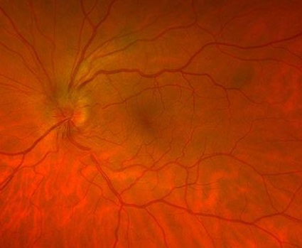
On exam there was a mild left APD; the vision was 20/20 in the right eye and 20/25 in the left eye. Fundus exam showed left superior optic nerve head swelling.
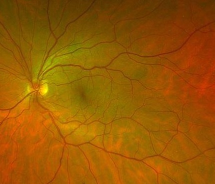
The patient was seen 3 months prior and was noted to have a crowded disc with c/d ratio less than 0.1 in both eyes. The superior optic disc
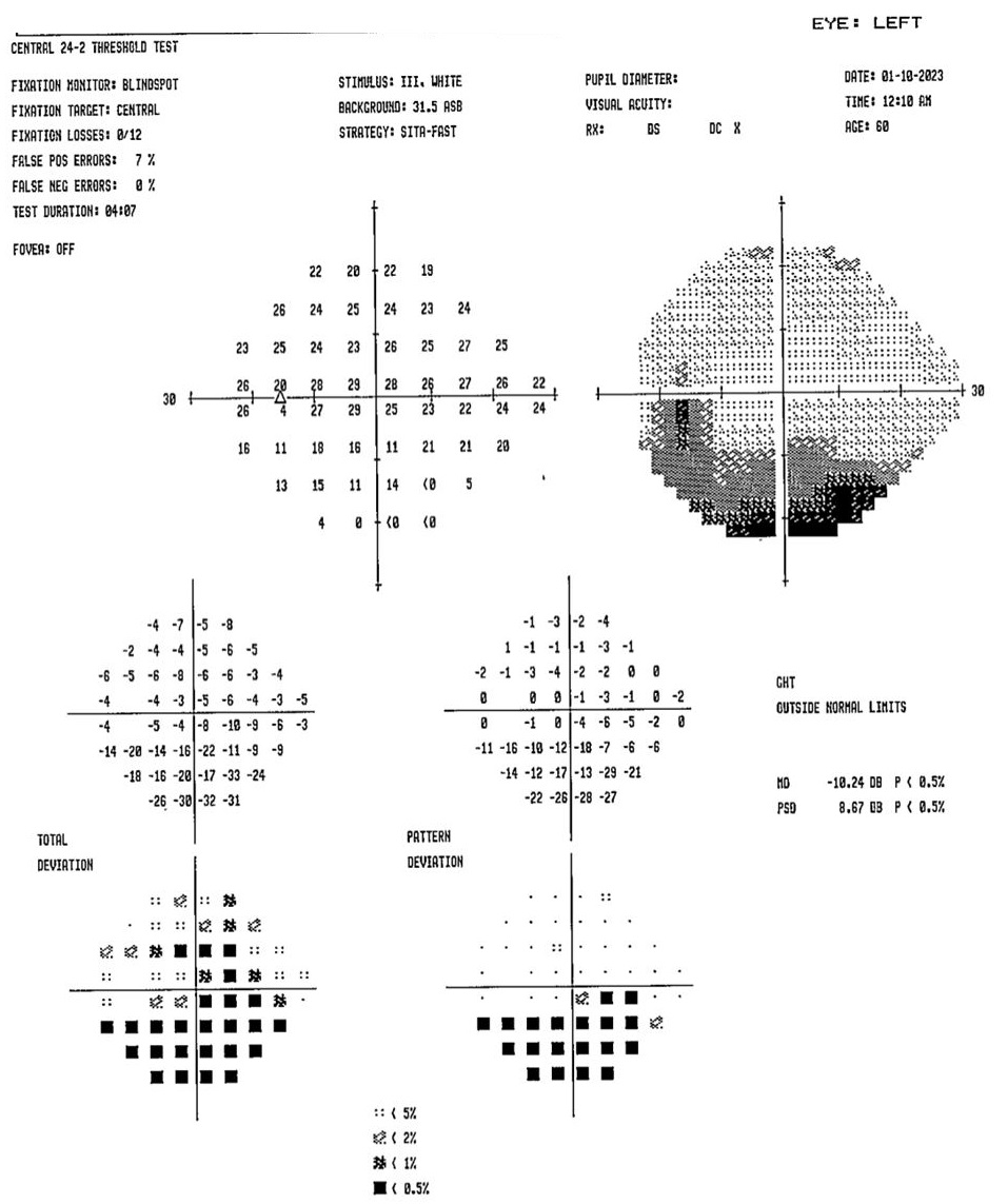
swelling seen clinically and on OCT RNFL corresponds to the inferior altitudinal defect seen on automated Humphrey visual field test (HVF 24-2).
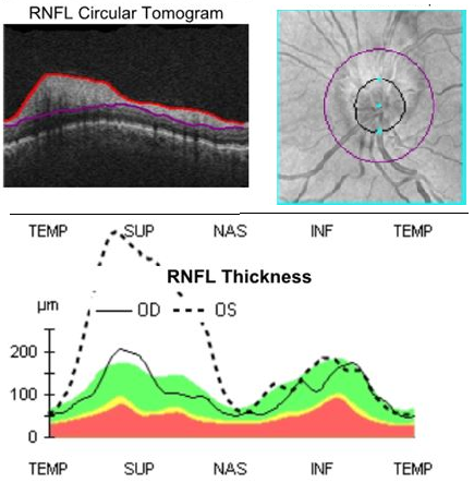
Full OCT RNFL of the right and left eye
Optos pseudocolor image on presentation
Optos pseudocolor image 3 months prior to presentation
Visual field of the left eye on presentation
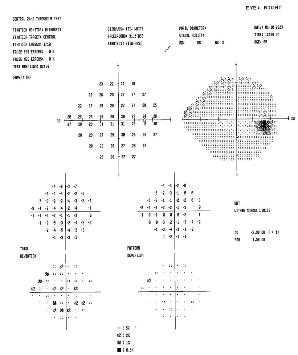
Visual field of the right (unaffected) eye on presentation